
Quantum Sensors in Medicine: Where They Already Have Practical Use
Quantum sensing has stopped being a purely laboratory story. In medicine, it now sits in a practical middle ground: a few technologies are already being used in real-world settings, several are in serious clinical pilots, and plenty are still waiting for the engineering and regulatory work that turns a clever measurement into a dependable medical tool. The common thread is sensitivity—quantum sensors can detect extremely weak magnetic or optical signals that conventional instruments either miss or can only capture with bulky, expensive infrastructure.
Wearable brain sensing with optically pumped magnetometers (OPM-MEG)
The most visible “already useful” area is magnetoencephalography (MEG) with optically pumped magnetometers. Traditional MEG uses superconducting sensors (SQUIDs) that require cryogenic cooling, making systems heavy, costly to run, and physically distant from the scalp. OPM-based systems work at or near room temperature and can be arranged as wearable arrays that sit closer to the head, which improves signal capture and makes movement-tolerant recordings more realistic in practice.
In 2026, OPM-MEG is widely discussed as a route to more accessible neuromagnetic measurements—especially in groups that are difficult to scan with classic systems, such as children or patients who cannot keep completely still. In clinics, the near-term value is not “replacing MRI” or “diagnosing everything”, but improving functional mapping and monitoring: identifying where epileptic activity begins, supporting presurgical planning, and tracking brain responses during tasks in a way that complements EEG.
What matters for hospitals is workflow reliability: shielding, sensor calibration, repeatability, and integration with existing neurodiagnostic pathways. The strongest centres treat OPM-MEG as an advanced extension of MEG rather than a magic new category—useful when timing and localisation of brain activity are the key clinical questions, and less relevant when structural imaging is the priority.
Where OPM-MEG helps diagnosis and monitoring right now
Epilepsy pathways are the clearest example. MEG is already used in specialised centres to localise epileptiform activity and guide surgical decisions, and OPM systems aim to deliver comparable outputs with potentially better comfort and flexibility. The clinical promise is practical: higher-quality data in patients who struggle with rigid scans, and the possibility of longer or repeated recordings for monitoring changes over time.
Paediatric and developmental neurology is another practical driver. A wearable arrangement can support recordings that would be difficult with a fixed helmet and strict “don’t move” rules. For clinicians, the benefit is not only comfort; it is the chance to gather interpretable data in real children rather than idealised conditions, which can reduce the gap between research-grade measurements and clinical reality.
There is also growing interest in treatment monitoring and neurorehabilitation research—tracking how brain networks respond to therapy, stimulation, or learning. That is not yet a standard hospital service everywhere, but it is a realistic use case because it aligns with what MEG is already good at: measuring functional dynamics with millisecond timing, then comparing patterns across sessions.
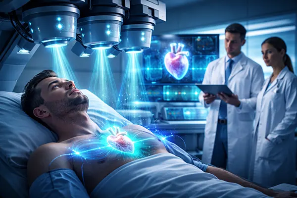
Quantum magnetometry for the heart and peripheral physiology
If the brain story is “specialist clinical use expanding”, the heart story is “early stage but compelling”. The heart produces magnetic fields as well as electrical signals. ECG is excellent for many purposes, but magnetic measurements can add a different view, potentially helping in situations where electrical recordings are ambiguous or where spatial mapping of activity is valuable.
Room-temperature quantum magnetometers are central here. Two families are most discussed: optically pumped magnetometers (also used for MEG) and diamond nitrogen-vacancy (NV) sensors. NV sensors use defects in diamond whose quantum properties change with magnetic fields; with suitable optics and microwaves, the readout becomes a very sensitive magnetometer in a compact form factor.
In 2026, you should treat quantum-based magnetocardiography (MCG) as an emerging clinical candidate rather than an established routine test. The practical work is happening—engineering demonstrations, pilot measurements, and company-led development—but the clinical “product” is still being shaped: standardising protocols, proving diagnostic value against ECG plus imaging, and building systems that work outside ultra-controlled labs.
Practical pathways: from pilot MCG to monitoring applications
The near-term clinical niche for MCG is likely to be targeted, not universal. Think “add-on measurement when timing and spatial patterns of cardiac activation matter”, rather than a replacement for ECG. For example, non-invasive mapping could become useful in complex arrhythmias, therapy planning, or when clinicians want an additional signal channel without introducing catheters.
Another plausible pathway is bedside monitoring in specialist settings. Quantum sensors are attractive when they can deliver sensitive measurements with low maintenance burdens. If systems can be made compact, robust to environmental noise, and safe in crowded wards, they could support continuous or repeated monitoring in high-risk cardiac patients—provided clinical trials show that the extra signal changes decisions or outcomes.
It is also worth watching peripheral applications where tiny magnetic signals matter—such as muscle activity or nerve function—because the same sensor families can, in principle, detect biomagnetic fields beyond the heart and brain. The clinical logic is similar: they become worthwhile when they answer a question that electrical sensors, ultrasound, or standard imaging cannot answer cleanly.
Quantum sensors in labs and imaging: real utility, but not always bedside-ready
Not all medical quantum sensing is about biomagnetism. Some of the most practical uses today sit in laboratory and imaging-adjacent workflows. Here, the “quantum” part often means improved sensitivity at low signal levels—useful for reducing dose, improving contrast, or measuring microenvironments that are hard to probe otherwise.
A concrete example is nanodiamond sensing in biomedical research. NV centres inside nanodiamonds can act as tiny probes that report on magnetic fields, temperature, or local chemistry at microscopic scales. In 2026, this is more commonly a research and translational tool than a routine diagnostic test, but it is already practical in the sense that labs can deploy it to study cell environments, drug effects, or micro-scale thermal changes that are relevant to disease processes.
On the imaging side, quantum optical methods—such as squeezed-light approaches and other quantum-enhanced measurements—are being explored to improve signal-to-noise at lower illumination levels. The practical medical angle is safety and fidelity: achieving clearer images while minimising light exposure in delicate tissue, or improving sensitivity in modalities related to optical coherence techniques. The honest status is mixed: some methods are impressive in controlled demonstrations, but broad clinical adoption depends on making them rugged, affordable, and easy to maintain.
How to judge “practical” in 2026: evidence, integration, and limits
For a hospital or clinic, “practical” is less about headlines and more about the chain from measurement to decision. A quantum sensor becomes clinically meaningful when it produces a repeatable output, has clear reference ranges or interpretation rules, and fits into existing diagnostic routes. That includes basics: calibration routines, quality control, staff training, and compatibility with other equipment.
Evidence needs to be specific. The best studies compare quantum-enabled measurements against current standards in real patient cohorts, not only against idealised lab benchmarks. For neuromagnetic applications, that means showing reliable localisation and test–retest stability. For cardiac or lab applications, it means demonstrating that the sensor changes diagnoses, treatment plans, or monitoring decisions—and does so consistently.
Finally, limitations should be stated plainly. Biomagnetic systems often require careful control of magnetic noise; wearable designs can introduce motion artefacts; and advanced optical methods can be sensitive to alignment, temperature drift, and component ageing. In 2026, the sensible view is optimistic but grounded: quantum sensors are already useful in certain specialist contexts, and the next step is not discovering new physics, but building dependable medical systems around what already works.